M paper bits
- 4.2
- Scaling
- Standardization outperformed Normalization (Fig. XX) to an extreme degree, therefore only results for Standardization will be reported in the following sections.
- SW and Size
- Fig. XX shows results with and without sample weights, and on the same axis (but as separate experiment) different patch sizes. A larger patch size has a positive effect on scores (results in line with previous research [Tkachenko et. al]), but require more resources, with a patch size of 7 already being impractical to experiment on.
- Smoothing
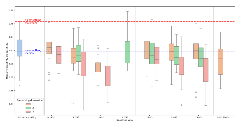
- Given the large amount of combinations for the various smoothing parameters tested, only part of them could be selected for further evaluation.
- Our criteria for inclusion was a meaningful improvement of the median or of the maximum achieved score over the no-smoothing baseline in any of the dimensions.
- Fig. XX shows the results without smoothing applied (blue), and all tested combinations.
- Smoothing, in most cases, has a negative effect on the scores, with the median decreasing in all but two instances (1D MF-7 and 1D GF-0.5), and the maximum achieved value being surpassed only by 2D GF-2 and 2D MF-3. This is not consistent with previous research (Tkachenko et al.) that showed improvements from the application of MF.
- As per our criteria, we included only GF (0.5 and 2) in our test, as they improved the median and maximum scores respectively by a meaningful margin.
- Scaling
Introduction
Cancer diseases are one of the leading causes of death worldwide1. Colorectal cancer — third by incidence and second by mortality2 — accounts for a significant portion of cancer-related morbidity. Techniques usable during preoperative/diagnostic or intra/post-operative stages (e.g. to detect cancer regions and ensure tumor-free resection margins) can aid in cancer diagnosis and improve outcomes.
Hyperspectral imaging (HSI) can be seen as combination of a digital camera with a spectrometer, creating image data in the shape of hypercubes: two spatial dimensions and the third, containing the spectrum for each pixel. It has shown potential in discriminating between tissue structures by analyzing their interaction with light, and thereby distinguish healthy and pathological (e.g. cancerous) tissue in a contactless, radiation-free and non-invasive manner, and has seen increasing adoption in the medical field. 3
HSI has been used for cancer detection in humans, from skin (cit) to breast (cit) to oral (cit) cancer, as well as colorectal cancer (cit). The accuracy depends on the approaches used to classify and analyze the hyperspectral image data, with Machine Learning (ML) and especially Deep Learning (DL) approaches being favoured in recent years 4.
Cancer detection with HSI and DL presents challenges that start at the dataset aquisition stage: hyperspectral imaging and ground truth labeling requires devices and medical expertise, leading to smaller datasets than usually found in non-medical domains.
HSI images themselves present many inherent difficulties due to their complexity5, and in the medical domain can include class imbalances, light/blood reflections and variations in tissue structure, all presenting challenges in developing accurate and robust models.
Previous studies have explored the impact of pre- and post-processing steps on the supervised classification of cancer in hyperspectral images, highlighting the importance of optimized data processing techniques 6 7 8.
This study builds upon these findings by addressing the challenge of optimizing the preprocessing and modeling stages of HSI data analysis for cancer diagnostics. The primary objective is to identify the most influential preprocessing techniques and model parameters to enhance the predictive accuracy of machine learning models. By systematically exploring various combinations of preprocessing steps, such as scaling, smoothing, filtering, class weighting (cancerous tissue is less represented) and sample weighting (to increase model sensitivity to smaller areas).
Testing all parameters would have been prohibitively difficult. To overcome these challenges this paper introduces an iterative tuning pipeline designed to optimize preprocessing combinations and model parameters systematically. The pipeline allows for testing multiple combinations on a representative subset of the dataset before applying the best-performing methods to the entire dataset.
- Improve
-
Concrete DL and bit
- More examples incl. more recent
-
smaller sizes ds concretize
-
M sent paper from Marianne about blood and reflections
-
no one does blood — we do
-
no one does smoothnig — we do
-
end/description:
- what made it awesome
-
- complexity becasue of small dataset
- weight initialization and care so that few confounding this
- reproducible and вдумчтв
- один порядок та же точка без рендома
-
- smoothing and bg extraction tested, and there’s little smoothing
-
- what made it awesome
-
-
Torre, Lindsey A., Rebecca L. Siegel, Elizabeth M. Ward, and Ahmedin Jemal. “Global Cancer Incidence and Mortality Rates and Trends—An Update.” Cancer Epidemiology, Biomarkers & Prevention 25, no. 1 (January 1, 2016): 16–27. https://doi.org/10.1158/1055-9965.EPI-15-0578. ↩︎
-
Sung, Hyuna, Jacques Ferlay, Rebecca L. Siegel, Mathieu Laversanne, Isabelle Soerjomataram, Ahmedin Jemal, and Freddie Bray. “Global Cancer Statistics 2020: GLOBOCAN Estimates of Incidence and Mortality Worldwide for 36 Cancers in 185 Countries.” CA: A Cancer Journal for Clinicians 71, no. 3 (May 2021): 209–49. https://doi.org/10.3322/caac.21660. ↩︎
-
Barberio, Manuel, Sara Benedicenti, Margherita Pizzicannella, Eric Felli, Toby Collins, Boris Jansen-Winkeln, Jacques Marescaux, Massimo Giuseppe Viola, and Michele Diana. “Intraoperative Guidance Using Hyperspectral Imaging: A Review for Surgeons.” Diagnostics 11, no. 11 (November 8, 2021): 2066. https://doi.org/10.3390/diagnostics11112066. ↩︎
-
Oswald, Willaim, Craig Browning, Ruthba Yasmin, Joshua Deal, Thomas C. Rich, Silas J. Leavesley, and Na Gong. “Fluorescence Excitation-Scanning Hyperspectral Imaging with Scalable 2D–3D Deep Learning Framework for Colorectal Cancer Detection.” Scientific Reports 14, no. 1 (June 26, 2024): 14790. https://doi.org/10.1038/s41598-024-64917-5. ↩︎
-
AL-Alimi, Dalal, Mohammed A.A. Al-qaness, Zhihua Cai, and Eman Ahmed Alawamy. “IDA: Improving Distribution Analysis for Reducing Data Complexity and Dimensionality in Hyperspectral Images.” Pattern Recognition 134 (February 2023): 109096. https://doi.org/10.1016/j.patcog.2022.109096. ↩︎
-
Aboughaleb, Ibrahim H., Mohamed Hisham Aref, and Yasser H. El-Sharkawy. “Hyperspectral Imaging for Diagnosis and Detection of Ex-Vivo Breast Cancer.” Photodiagnosis and Photodynamic Therapy 31 (September 2020): 101922. https://doi.org/10.1016/j.pdpdt.2020.101922. ↩︎
-
Tkachenko, Mariia, Claire Chalopin, Boris Jansen-Winkeln, Thomas Neumuth, Ines Gockel, and Marianne Maktabi. “Impact of Pre- and Post-Processing Steps for Supervised Classification of Colorectal Cancer in Hyperspectral Images.” Cancers 15, no. 7 (April 5, 2023): 2157. https://doi.org/10.3390/cancers15072157. ↩︎
-
Aloupogianni, Eleni, Masahiro Ishikawa, Naoki Kobayashi, and Takashi Obi. “Hyperspectral and Multispectral Image Processing for Gross-Level Tumor Detection in Skin Lesions: A Systematic Review.” Journal of Biomedical Optics 27, no. 06 (June 8, 2022). https://doi.org/10.1117/1.JBO.27.6.060901. ↩︎